
DNA Extraction and Metagenome Sequencing in Microbiology
Estimates state, that only 1-2% of all microorganisms on the planet have been cultivated in a laboratory. This means, that the vast majority of bacteria and archaeacannot be surveyed by culture, and DNA is often the only reliable source of information about which microbes are present in a sample, what they can do and how many there are. This article covers where microbial DNA is found, how bacteria and archaea differ at the molecular level, how DNA is extracted, and which sequencing and quantification methods turn a sample into usable metagenome data. It also explains where bias enters the process.
Table of Contents:
What is DNA and what does it encode in a microbe?
Where do you find microbial DNA from living cells?
What can DNA from dead cells reveal about past microbes?
How does DNA differ between bacteria and archaea?
How can DNA be extracted from microorganisms?
Which sequencing methods identify the microbes in a DNA sample?
How is DNA quantified with qPCR and digital PCR?
What are the main pitfalls and biases in DNA analysis?
Why does careful DNA analysis matter?
What is DNA and what does it encode in a microbe?
Deoxyribonucleic acid is a long molecule built from two strands that wind around each other in a double helix. Each strand is a chain of sugar and phosphate units, and attached to each sugar is one of four bases: adenine (A), thymine (T), guanine (G) and cytosine (C). A always pairs with T, and G always pairs with C, which lets a cell copy its genome accurately before it divides.
The order of these bases is the code. Stretches of the sequence form genes that carry instructions for proteins, such as the enzymes a methanogen needs to produce methane or a sulfate reducer needs to generate hydrogen sulfide. Determining that order is called sequencing, and the full set of genes from one organism is its genome. When scientists sequence the genetic material of an entire community at once, the result is a metagenome: a combined catalog of every genome present in the sample, from the dominant species down to rare members. Comparing one metagenome with another, for example before and after a change in operating conditions, shows how a community responds.

Where do you find microbial DNA from living cells?
Microbes occupy almost every environment on Earth, and their genetic material goes wherever they go. Soil, lakes and oceans hold enormous microbial populations, as do the human gut and the surfaces of plants. Less obvious habitats matter just as much for industry. Porous rock kilometers below the surface, formation water in underground gas storage sites, cooling water circuits, geothermal brines and the inner walls of pipelines all host active communities, often organized as biofilms.
Some of these sites are extreme. Salt-saturated brines are home to halophiles, while hot reservoirs favor heat-loving archaea. Many deep habitats are also anaerobic and under high pressure, which makes collecting cells without disturbing them a technical challenge that dedicated high-pressure sampling methods are designed to solve.
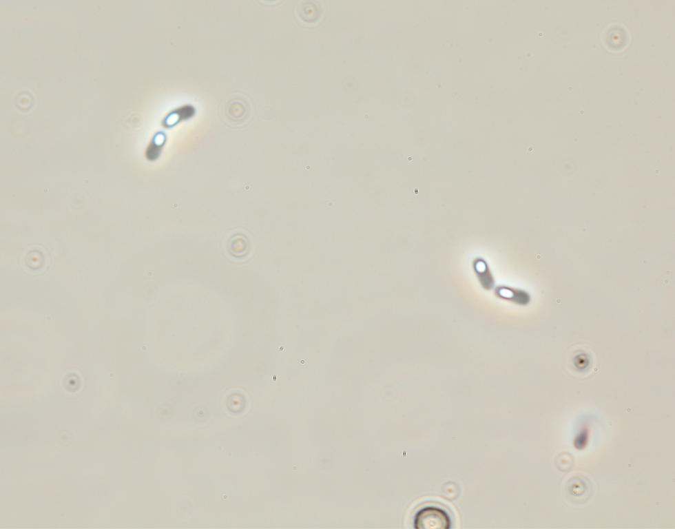
Using a slow-decompression technique, the microbial cells are kept intact and alive. This results are even visible within the samples.

What can DNA from dead cells reveal about past microbes?
Not every gene fragment in a sample belongs to a living organism. When cells die, their genetic material is either released into the surroundings or stays trapped inside intact but inactive cells. This relic or extracellular DNA can persist for days to thousands of years, depending on temperature, enzymatic activity, and even mineral surfaces. Clay particles in particular bind it and protect it from degradation.
Relic material is useful and problematic at the same time. Permafrost, lake sediments and ice cores preserve genetic traces that let researchers reconstruct which microbes lived there long ago. In an active industrial system, however, the same leftovers blur the picture, because sequencing alone cannot tell living and dead cells apart. A metagenome from soil or sediment may include a substantial share of sequences from cells that are no longer alive, with one study estimating around 40 percent in soils. Treatments with propidium monoazide (PMA), which chemically masks genetic material from membrane-damaged cells before amplification, help separate living from dead signals in the final metagenome.
How does DNA differ between bacteria and archaea?
At the chemical level, it does not. The same four bases and the same double helix are found in plants, fungi, humans and every microorganism. The differences lie in how the molecule is packed, how its bases are balanced and how hard it is to reach.
Packaging: bacteria compact their DNA into a nucleoid using nucleoid-associated proteins such as HU (heat-unstable nucleoid protein) and H-NS (histone-like nucleoid-structuring protein), while many archaea wrap theirs around histone proteins related to those in human cells.
Base composition: the share of G and C bases in DNA, known as GC content, ranges from below 20 percent to above 70 percent across microbes, and many haloarchaea sit at the high end.
Accessibility: the cell envelope decides how easily DNA can be released, and this is where the two domains differ most.
Bacterial cell walls are built from peptidoglycan, which enzymes such as lysozyme can break down. Archaea have no peptidoglycan at all. Methanogens such as Methanobacterium and Methanothermobacter use pseudomurein, which resists lysozyme, while Methanosarcina can form thick polysaccharide layers around cell clusters. Some haloarchaea, by contrast, burst open as soon as the surrounding salt is removed. These contrasts directly affect how much of each group ends up in a metagenome after sequencing.

How can DNA be extracted from microorganisms?
Every extraction follows the same logic: open the cells, release the genetic material and separate it from proteins, membranes, salts and other contaminants. Labs combine three types of approaches.
Chemical lysis uses detergents such as SDS, chaotropic salts and enzymes like lysozyme or proteinase K to dissolve cell envelopes and free the DNA.
Physical lysis breaks cells mechanically by bead beating, freeze-thaw cycles or sonication, which opens even tough cells but fragments the DNA in the process.
Purification with phenol-chloroform, silica columns or magnetic beads removes inhibitors and leaves DNA ready for analysis.
The central trade-off is length versus yield. Vigorous bead beating breaks open the widest range of microbes and delivers high amounts, but the genetic material comes out in short pieces. Gentle enzymatic protocols preserve the long, intact molecules that long-read sequencing needs, yet they may leave hardy cells such as pseudomurein-walled methanogens unopened. Commercial kits are fast and reproducible, though they can perform poorly with low-biomass samples and carry trace contaminants that appear as false signals in a sparse metagenome. The right choice depends on whether the goal is a broad metagenome survey, a complete genome or a sensitive count of one target.
Which sequencing methods identify the microbes in a DNA sample?
Qualitative analysis answers two questions: who is there and what they are capable of. Sequencing technology has moved through three generations. Sanger sequencing, developed in the 1970s, reads one fragment at a time with high accuracy and is still used to confirm single genes or pure cultures. Short-read platforms such as Illumina sequencing read millions of fragments in parallel, typically 150 to 300 bases long, which made large community studies affordable. Long-read technologies, including PacBio and Nanopore sequencing, produce reads of many thousands of bases, which helps assemble complete genomes from a complex metagenome.
Two targets dominate microbial work. 16S profiling amplifies and reads a single marker gene, the 16S rRNA gene, which every bacterium and archaeon carries. It is cost-effective and shows community composition, usually down to genus level. Metagenome sequencing, often called shotgun sequencing, reads all the genetic material in a sample without amplifying one target first. A metagenome reveals not only which organisms are present but also which metabolic genes they carry, and with enough depth it yields metagenome-assembled genomes of species that have never been cultured. Sequencing depth, meaning the number of reads per sample, decides how many rare members show up in each metagenome.
How is DNA quantified with qPCR and digital PCR?
Knowing that a sulfate reducer is present is not the same as knowing whether there are ten cells or ten million. Sequencing data only show proportions, so absolute numbers require targeted quantification alongside metagenome sequencing.
Quantitative PCR (qPCR) tracks the amplification of a chosen gene in real time using fluorescence. The earlier the signal appears, the more copies the sample contained, and a standard curve converts that signal into copy numbers. Digital PCR splits the reaction into thousands of tiny partitions, counts how many turn positive and calculates absolute copy numbers without a standard curve. It also tolerates inhibitors better, which helps with oily or saline field samples.
Typical targets include total bacterial or archaeal 16S rRNA genes and functional genes such as dsrA for sulfate reducers or mcrA for methanogens. In practice, qPCR and digital PCR complement sequencing: the metagenome shows which groups matter, and targeted assays track how their numbers change over weeks or months.

What are the main pitfalls and biases in DNA analysis?
Each step between sampling and final data can distort the result, and errors early in the chain carry through to the end. A metagenome is only as representative as the sample behind it.
Sample quality: oxygen exposure, pressure loss, delays or warm storage change the community or degrade its DNA before analysis starts.
Extraction: methods that miss tough cells leave their DNA behind and underrepresent those groups in the metagenome.
Primer design: so-called universal primers match some lineages poorly, so DNA from mismatched groups amplifies weakly or not at all before sequencing even begins.
Sequencing method: short reads struggle with repetitive regions and extreme GC content, while long reads have historically carried higher error rates, and each platform skews DNA data in its own way.
Archaea: tough envelopes, divergent 16S sequences and thin coverage in reference databases make archaeal DNA especially easy to underestimate.
Gene copy numbers add another layer. Bacteria carry between one and about 15 copies of the 16S rRNA gene per genome, so high copy counts do not always mean high cell counts. Archaea need particular attention, since methanogens and other archaea play central roles in methane formation and corrosion in storage and energy systems. A protocol tuned for bacteria can make them nearly invisible. Archaea-specific primers, adapted lysis steps and cross-checks between 16S profiling and metagenome sequencing are the most reliable safeguards.
Why does careful DNA analysis matter?
Microbial genetic material holds detailed information about who lives in a system and what they are doing, but only when it is sampled, extracted and analyzed with its biases in mind. Combining 16S profiling, metagenome sequencing and targeted quantification gives the most complete picture, especially for archaea that standard workflows tend to miss.

Comments