
Inside the Bacterial Cell: Walls, Spores and Surface Structures
In 1884, the Danish physician Hans Christian Gram flooded a slide of bacteria with a violet dye, rinsed it with alcohol and noticed that some cells stayed purple while others lost their color completely. He had stumbled on one of the deepest structural divides in the bacterial world. More than a century later, the Gram stain is still one of the first tests run on an unknown isolate, because the color a cell takes tells you a great deal about how it is built. This article walks through that build: the basic parts every bacterium shares, the two main types of cell wall, and the specialized structures that let bacteria survive, move, stick and trade with their surroundings.
The basic bacterial cell
A typical bacterium is only one to a few micrometers long, and it has no nucleus and no membrane-bound organelles. Everything happens inside a single compartment bounded by the cytoplasmic membrane. This membrane is a thin double layer of phospholipids packed with proteins. It controls what enters and leaves the cell, and because bacteria have no mitochondria, it is also where energy is generated. Respiratory enzymes in the membrane pump protons outward, and the flow of those protons back in drives the enzyme ATP synthase.
Inside the membrane lies the cytoplasm, a dense gel of water, enzymes, metabolites and ribosomes. The genetic material sits in a region called the nucleoid, usually as a single circular chromosome, often with smaller DNA circles called plasmids that carry extra genes such as antibiotic resistance.
Wrapped around the membrane is the cell wall. In bacteria, its core material is peptidoglycan, a mesh of sugar chains cross-linked by short peptides. The cytoplasm is full of dissolved molecules, so water constantly pushes inward and the cell builds up internal pressure. The wall is what stops it from bursting. It also gives the cell its shape, whether rod, sphere or spiral. Penicillin and related antibiotics work by blocking the enzymes that cross-link this mesh.
Gram-positive and Gram-negative cell walls
The Gram stain separates bacteria by how their walls are built, and the difference goes far beyond color.
Gram-positive bacteria have a thick peptidoglycan wall, roughly 20 to 80 nanometers across and made of many stacked layers. Threaded through it are teichoic acids, chain-like polymers that add negative charge and help anchor the wall. When the violet dye and iodine form a complex inside the cell, the thick wall traps it, and the alcohol rinse cannot wash it out. Bacillus, Clostridium and Staphylococcus belong to this group.
In Gram-negative bacteria, the design is more layered. The peptidoglycan is only a few nanometers thick and sits in a space called the periplasm. Outside it is a second, outer membrane, whose outer face is covered in lipopolysaccharide. The alcohol rinse disrupts this outer membrane, the dye escapes through the thin wall, and the cells take up the pink counterstain instead. Escherichia coli is the classic example. So is Desulfovibrio, a sulfate-reducing bacterium known for its role in corrosion.
The outer membrane matters well beyond the microscope. It acts as an extra barrier against detergents and many antibiotics, which is one reason Gram-negative infections can be harder to treat. Not every bacterium fits neatly into either group. Mycobacteria add a waxy coat of mycolic acids that resists the standard stain, and mycoplasmas have no wall at all.

Endospores
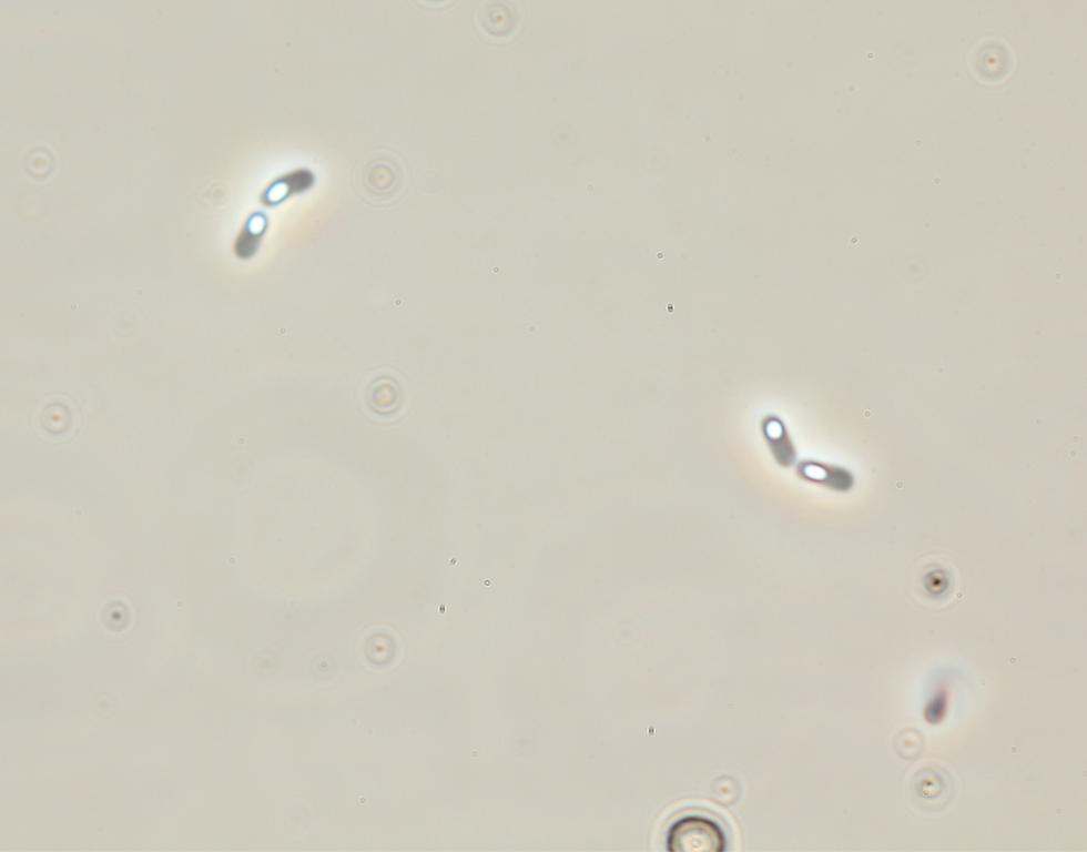
Some Gram-positive bacteria, most famously Bacillus and Clostridium, have an emergency strategy. When nutrients run out, the cell builds a copy of its chromosome into a dormant, heavily protected structure called an endospore. The spore core is strongly dehydrated and rich in calcium dipicolinate, and it is wrapped in several protective coats. Metabolism essentially stops.
Endospores tolerate boiling, drying, UV radiation and many disinfectants. Laboratory sterilization in an autoclave, at 121 °C under pressure for at least 15 minutes, is designed specifically to kill them. When conditions improve, a spore can germinate and grow back into an active cell, sometimes after many years.
Spore formers are common in anaerobic environments. Desulfotomaculum, a Gram-positive sulfate reducer, forms endospores, which helps it persist through periods of stress in industrial and subsurface systems. That persistence is worth keeping in mind whenever a system has been cleaned or treated and a problem returns.
Appendages for movement, attachment and exchange
Many bacteria carry thin protein structures that reach out from the cell surface.
Flagella are long, helical filaments driven by a rotary motor anchored in the cell envelope. The motor is powered by ions flowing across the membrane and can spin at hundreds of revolutions per second. By switching rotation direction, a bacterium alternates between swimming runs and random tumbles, which lets it move toward nutrients and away from harmful substances.
Fimbriae and pili are shorter, thinner filaments, often present in large numbers. Their main job is attachment to surfaces, host tissues or other cells. Attachment is the first step in forming a biofilm, the kind of structure that creates anaerobic niches in aerated industrial water systems. Some pili, known as type IV pili, can also extend and retract, pulling the cell along a surface in a jerky motion called twitching motility.
Conjugative pili connect two cells so that DNA, usually a plasmid, can pass from one to the other. This is how resistance genes spread between bacteria. It is gene exchange rather than reproduction. Bacteria multiply by binary fission, where one cell divides into two.
Nanowires are conductive filaments that let some bacteria pass electrons to solid surfaces outside the cell. Geobacter sulfurreducens uses them to breathe on iron oxide minerals. These filaments were first described as modified pili, while later work pointed to chains of cytochrome proteins instead. Electron transfer of this kind is also discussed as one route by which microbes interact with metal surfaces.
Surface layers and coatings
The outermost surface of a bacterium is where it meets the environment, and many species add one more layer there.
An S-layer is a two-dimensional crystal made of a single protein or glycoprotein repeated thousands of times. The subunits assemble on their own into a regular lattice that covers the entire cell like chain mail. S-layers are found in many bacteria and in most archaea. They protect against predators and enzymes, act as a molecular sieve with pores of uniform size, and help cells attach to surfaces. Because the lattice is so regular, S-layer proteins have also been studied as templates in nanotechnology.
Other bacteria surround themselves with a capsule or slime layer made mostly of polysaccharides. This sticky coat helps cells resist drying, hide from immune cells and hold together in biofilms. Capsules show up well under the microscope with simple negative staining, and our article on shapes, colors and communities of microorganisms covers more of what microscopy reveals.

A different design in archaea
Archaea look much like bacteria under the microscope, but their cell biology follows a completely different design. Their membranes are built from different lipids, their walls contain no peptidoglycan, and many rely on the S-layer alone. We will take a closer look at the archaeal cell in an upcoming article.


Comments